Virtual reality games are becoming increasingly popular. They allow users to fully immerse themselves in another, virtual world. Did you know that virtual reality systems are also used in research ? It is the case in the study of the brain and its mechanisms. This article is going to explain how it is possible to track neuronal activities in fish brains, using virtual reality.
Studies with zebrafish where virtual reality is involved
Virtual reality is used to study behavior and physiology for which feedback is important to the process. This is notably the case for locomotion and motor adaptation. In fact, zebrafish have to modulate and adapt their locomotion to the environment, being an important point for the survival of the fish. Locomotion processes in fish have to deal with changes in muscle temperature, fatigue, changes in water viscosity or changes in the wind (4).
Whole-brain activity is obtained by analyzing the relationship between spontaneous brain activity and spontaneous behavior (2). Virtual reality induces visual stimuli such as shoal images (5), prey imaging (6) or water flow (1). Virtual reality is widely used to study spatial cognition, social interaction or also collective behavior (7).
An interesting aspect is the brain reaction when visual feedback following a motor command does not meet expectation. In this situation, zebrafish can learn to adapt the strength of subsequent motor commands (1). With virtual reality, researchers were able to show that larval zebrafish apply a short-term learned change in sensorimotor transformation (1).
Another study shows that adult zebrafish prefer swimming with congeners than alone. When images of a shoal are presented to the zebrafish under virtual reality, he swims in the shoal direction (5).
The method
Virtual reality can be used with larvae or adult zebrafish. The advantages of using larvae are that they are transparent and can be restrained in agarose (5). In fact it is important to restrain the fish movement to be able to obtain performing images. Adults are more difficult to maintain because they can’t be immobilized in agarose.
But why is using adults in some studies interesting? Because in contrast to larvae, adults have a broader behavioral repertoire. Adult zebrafish are more appropriate in social interactions, innate behavior, learning and cognitive behavior studies (5).
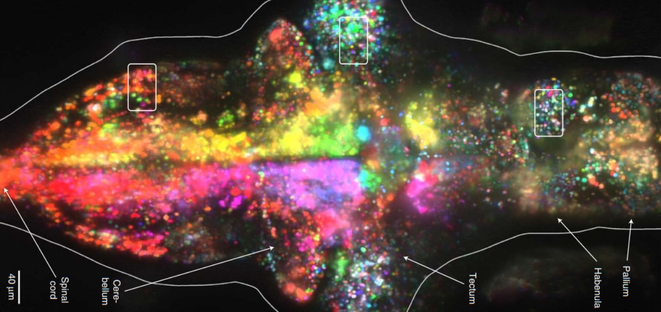
In vivo brain system visualization is achieved by using transgenic zebrafish expressing GCaMP2 (calcium indicator). This indicator allows the monitoring of a single cell neuron which can be seen by two photon microscopy (1,5). This method enables a high resolution functional neuronal recording (6).
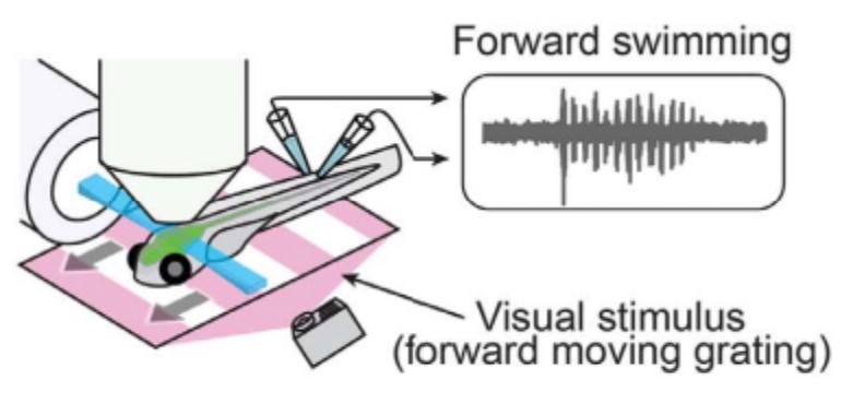
Then, brain monitoring can be completed by two other options. The first option is called Fictive swim and records the activity of a group of motor neuron axons from both sides of the tail to analyze its oscillations and locomotion (6). The name fictive swim came from the fact that fish are paralyzed and so they can’t really move. When a motor command is recorded, it is translated into visual feedback (1). The second option records tail movements with a high speed camera. In this experience, the zebrafish head is blocked but the rest of the body is free. The major advantage of this method is that it provides fine tracking of tail kinematics and proprioceptive feedback (6). Jouary and al. (2016) generated a library of larvae movements in free swimming conditions depending on larvae behavior repertoire to enable researchers to make a relationship between kinematics and change in orientation and position of the larvae (6).
Transgenic zebrafish are placed in a way to directly receive visual stimuli, as shown in figure 1.
With precise analysis and images, zebrafish have to be in good health and without malformations. This is necessary to be sure that movements and reactions recorded aren’t caused by a malformation. In this point, the use of the EggSorter can be really useful to avoid error and to ensure that the eggs are healthy and free from malformation or disease, or to select the transgenic line of interest.
Conclusion
Virtual reality has the same role in humans and zebrafish : simulating a situation. Using it with zebrafish and the current powerful technology allows the possibility to follow in vivo neuronal responses.
References
- Ahrens MB, Li JM, Orger MB, Robson DN, Schier AF, Engert F, et al. Brain-wide neuronal dynamics during motor adaptation in zebrafish. Nature. 2012 May;485(7399):471–7.
- Dunn TW, Mu Y, Narayan S, Randlett O, Naumann EA, Yang CT, et al. Brain-wide mapping of neural activity controlling zebrafish exploratory locomotion. eLife. 2016 Mar 22;5:e12741.
- Freeman J, Vladimirov N, Kawashima T, Mu Y, Sofroniew NJ, Bennett DV, et al. Mapping brain activity at scale with cluster computing. Nat Methods. 2014 Sep;11(9):941–50.
- Kawashima T, Zwart MF, Yang CT, Mensh BD, Ahrens MB. The Serotonergic System Tracks the Outcomes of Actions to Mediate Short-Term Motor Learning. Cell. 2016 Nov;167(4):933-946.e20.
- Huang KH, Rupprecht P, Frank T, Kawakami K, Bouwmeester T, Friedrich RW. A virtual reality system to analyze neural activity and behavior in adult zebrafish. Nat Methods. 2020 Mar;17(3):343–51.
- Jouary A, Haudrechy M, Candelier R, Sumbre G. A 2D virtual reality system for visual goal-driven navigation in zebrafish larvae. Sci Rep. 2016 Sep 23;6(1):34015.
- Stowers JR, Hofbauer M, Bastien R, Griessner J, Higgins P, Farooqui S, et al. Virtual reality for freely moving animals. Nat Methods. 2017 Oct;14(10):995–1002.
- Abdelfattah AS, Kawashima T, Singh A, Novak O, Liu H, Shuai Y, et al. Bright and photostable chemigenetic indicators for extended in vivo voltage imaging. Science. 2019 Aug 16;365(6454):699–704.
First image: Freeman J, Vladimirov N, Kawashima T, Mu Y, Sofroniew NJ, Bennett DV, et al. Mapping brain activity at scale with cluster computing. Nat Methods. 2014 Sep;11(9):941–50.