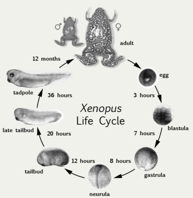
Xenopus laevis is the scientific name of the African clawed frog. This species is native from southern Africa and inhabits slow moving water or stagnant ponds. Its facility to adapt to different environments and conditions makes it considered as an invasive species in Europe, North America and South America (1). The consequence of its invasiveness is a decrease in the local biodiversity because of the competition with local amphibians and the fact that it eats local fishes and invertebrates (1). On the other hand, X.laevis is a very good model organism and is commonly used in life science research.
Xenopus laevis in research
Xenopus laevis is a model organism, particularly used to study ageing, human diseases, development, neurobiology and toxicology. It posses advantages such as (2) :
- 500-3'000 eggs per hatching
- Easy to maintain in laboratory
- Low cost
- Accessibility of the embryos
- Well known genome
- Possibility and ease to do transgenic individuals
Xenopus laevis share 79% of disease genes with humans (3). Disease groups identified are linked to the nervous, urinary, respiratory, cardiovascular and reproductive system. Cancer, genetic disease and physical disorder are also studied with these frogs.
The first discovery which involved Xenopus frogs allowed the conception, in 1938, of a pregnancy test. The test is based on changes in the hormonal rate in pregnant women and can detect pregnancy at early stages. When the pregnant woman’s urine is injected in the frogs, it triggers ovulation and the production of oocytes (4).
Since then, more than 30’000 articles have been written which relate to the use of Xenopus frogs (5).
The importance of sorting xenopus oocytes and embryos
Xenopus frogs have an external fertilization. The accessibility of the eggs is a major advantage because it allows manipulations that are not possible in mammals.
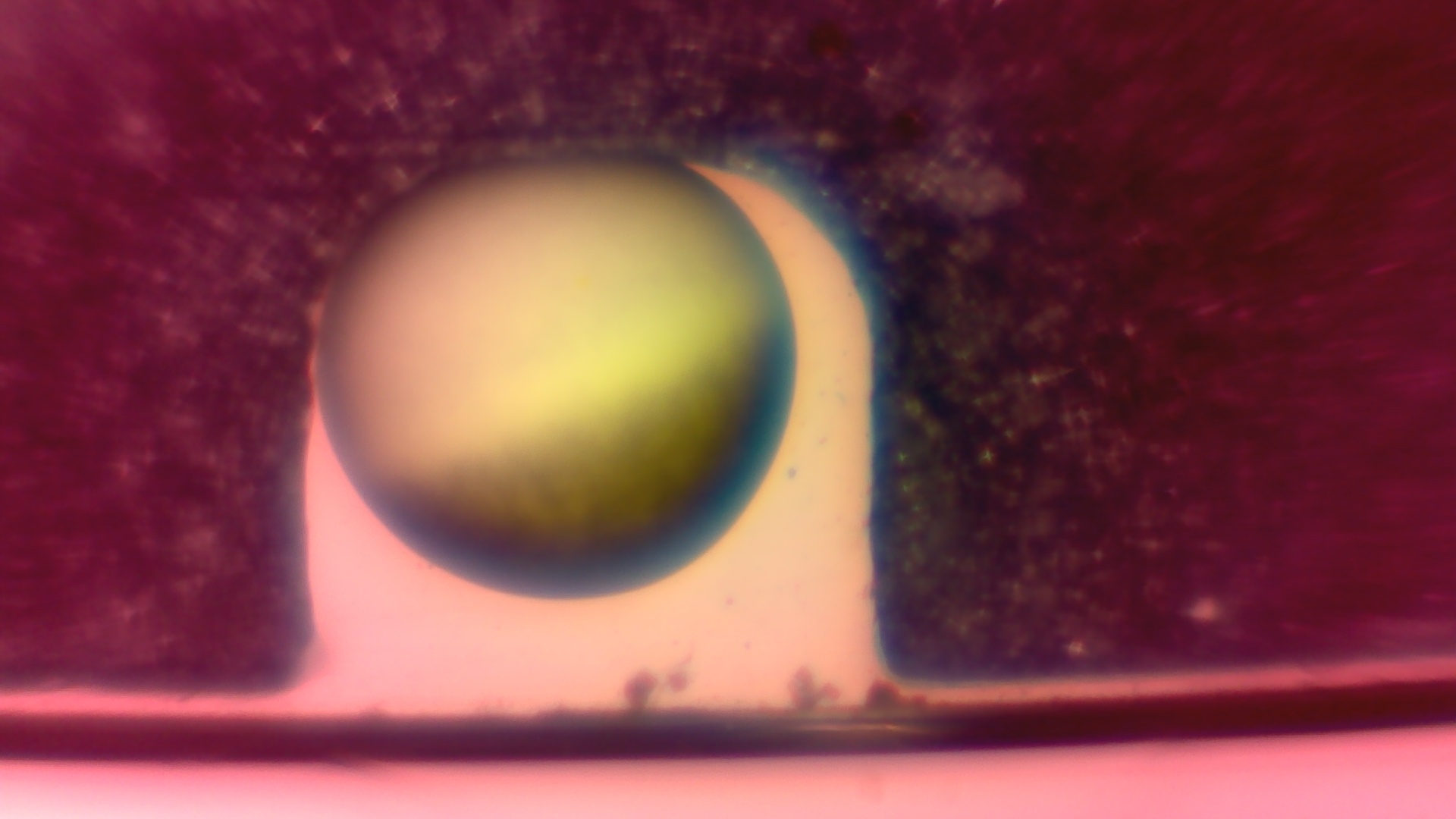
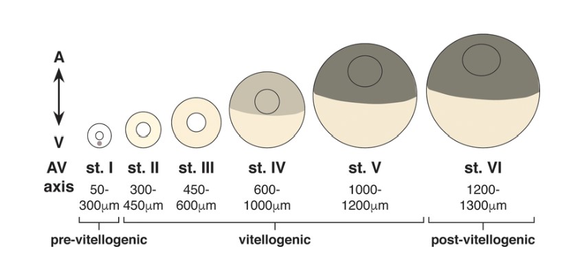
Oocytes are located in the ovarian cavity of female frogs. They represent the stage before the fertilisation. When oocytes are extracted from the ovarian tissue, all stages of the oogenesis are present but, normally, only the latest stages are used for the experiments. So firstly, it is necessary to sort the oocytes to have only the stages V and stages VI. After this, a second sorting is necessary to select only healthy oocytes.
Oocytes are often used to study single-cell expression systems (7). Oocytes contain a big number of proteins and transporters and, in this way, they are perfect to explore signal transduction pathways and cell fate (7,8).
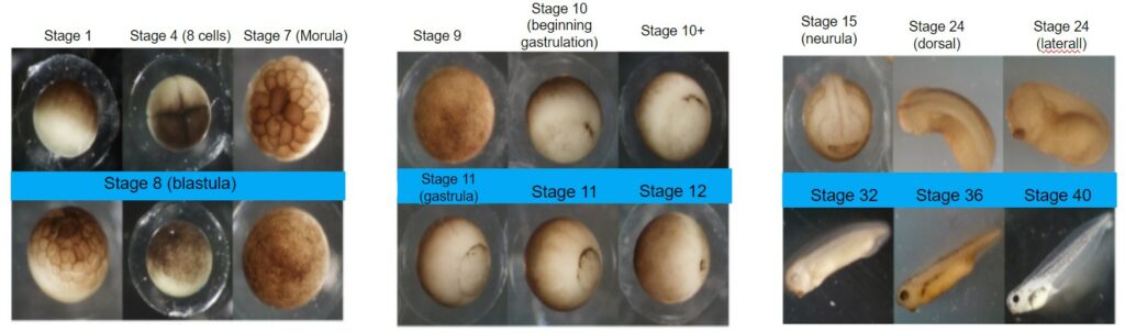
On the other hand, embryos are fertilized eggs. After the external fertilization, healthy embryos have to be sorted and all dying embryos ,with anomalies or unfertilized have to be removed. The next day, a second sorting is necessary to remove dead or abnormal embryos unseen the previous day.
Embryos are used to study different pathways and the development of some systems such as the neural system.
The EggSorter to automate the sorting
Manual sorting of oocytes is required to check the spherical shape, the brown pole which needs to be completely brown, without spots on it, and the equatorial line which should be well defined.
For the embryos, the sorting is made in two days. The first day, researchers have to remove all white eggs. These embryos are dead. Malformations can also be detected by the observations of the divisions. If an asymmetric division is present, the embryo is considered malformed. The second day, the same observations are made.
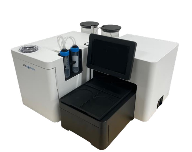
Aware that this manual handling and sorting takes up a lot of researchers’ time, Bionomous has modified the EggSorter, originally created to sort zebrafish eggs, so that it can also handle xenopus eggs. The EggSorter developed by our company automatically screens, sorts and dispenses small biological entities ranging in size from 0.5 to 2 mm. The EggSorter has been adapted to sort Xenopus laevis oocytes and embryos thanks to the addition of a tilting box, a reflective illumination system and a second camera with a focus on both sides.
Conclusion
Xenopus laevis appeared in recent years as a powerful model organism. But a major disadvantage is the time that the sorting of oocytes or embryos takes. With the EggSorter, 80% of the time is released. This automated process ensures more than 95% accuracy. If you want to learn more about the EggSorter and how it works or to have a demo, do not hesitate to visit our website and to contact us!
References
- Where Do Our Aquatic Models Come From ? – Bionomous [Internet]. [cited 2023 Aug 18]. Available from: https://bionomous.ch/articles/ecology-aquatic-models/
- Introducing Xenopus! – Bionomous [Internet]. [cited 2023 Aug 25]. Available from: https://bionomous.ch/articles/xenopus-research/
- Nenni MJ, Fisher ME, James-Zorn C, Pells TJ, Ponferrada V, Chu S, et al. Xenbase: Facilitating the Use of Xenopus to Model Human Disease. Front Physiol. 2019 Feb 26;10:154.
- Elkan ER. The Xenopus Pregnancy Test. Br Med J. 1938 Dec 17;2(4067):1253-1274.2.
- PubMed [Internet]. [cited 2023 Aug 26]. PubMed. Available from: https://pubmed.ncbi.nlm.nih.gov/
- Introduction to Xenopus – Xenbase [Internet]. [cited 2023 Aug 18]. Available from: https://www.xenbase.org/xenbase/anatomy/intro.do
- Yan Q, editor. Membrane Transporters in Drug Discovery and Development: Methods and Protocols [Internet]. Totowa, NJ: Humana Press; 2010 [cited 2023 Aug 25]. (Methods in Molecular Biology; vol. 637). Available from: http://link.springer.com/10.1007/978-1-60761-700-6
- Ferrell JE, Machleder EM. The Biochemical Basis of an All-or-None Cell Fate Switch in Xenopus Oocytes. Science. 1998 May 8;280(5365):895–8.
- https://sites.millersville.edu/jcebrathomas/cebra_thomas/DB_lab/Frog/frog_staging.html
- Mowry KL. Using the Xenopus Oocyte Toolbox. Cold Spring Harb Protoc. 2020 Apr 1;2020(4):095844. doi: 10.1101/pdb.top095844.