
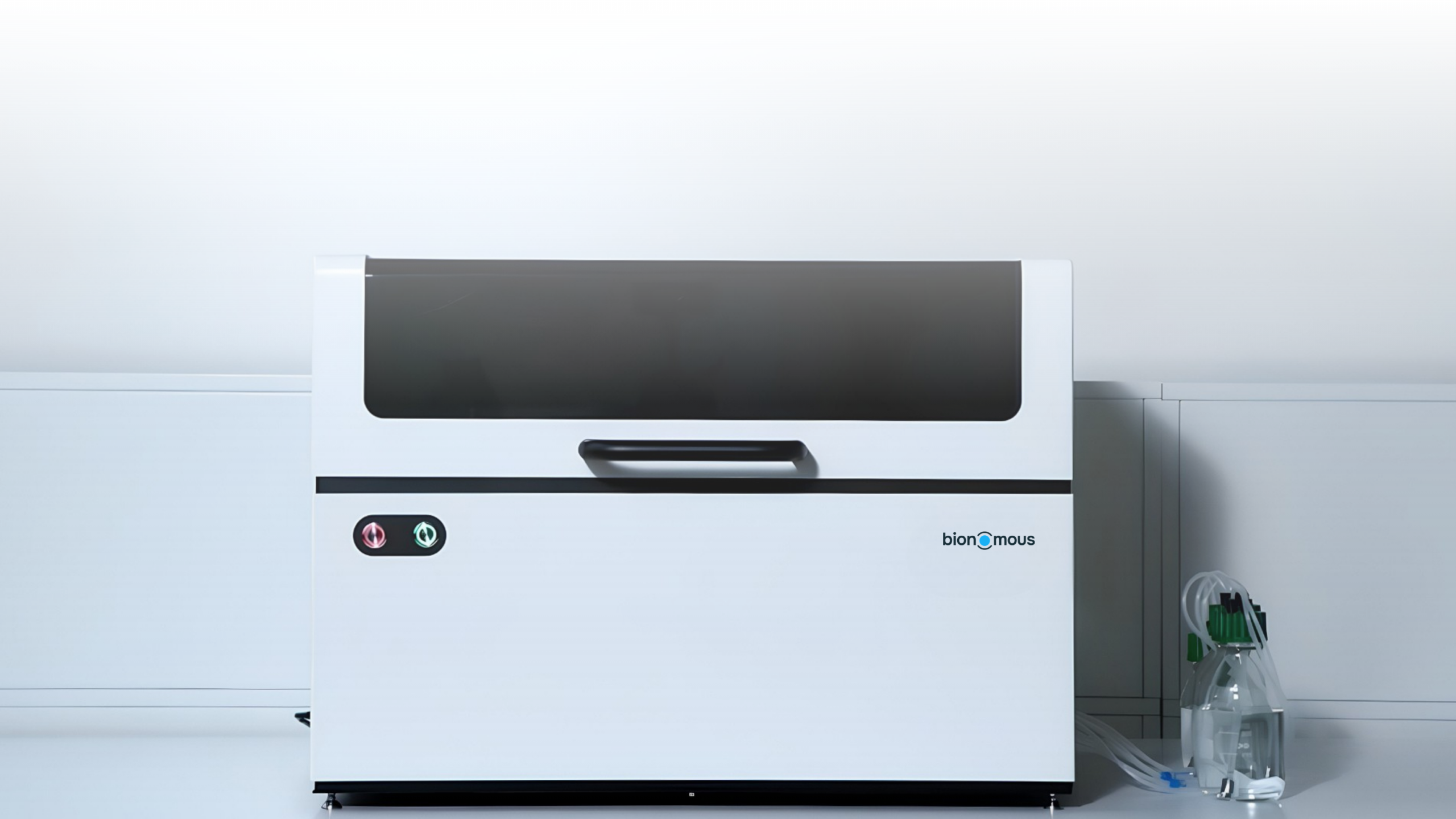
Zebrafish larvae have become one of the most versatile vertebrate models in biomedical research, from drug discovery to toxicology. Yet, before every screening experiment is a step that researchers rarely talk about: selection and distribution of the larvae. This month, we look at why sorting remains a bottleneck in zebrafish workflows, and how Sortivo turns this time-consuming step into a quick and automated routine.
Table of Contents
Introduction
Over the past decades, the zebrafish embryo has become a prominent model for studying gene function, human disease and drug response (1, 2, 3). It offers a combination of desirable features: low maintenance and husbandry costs, high fecundity, genetic diversity, physiology similar to that of traditional biomedical models, and reduced animal welfare concerns (4). It also shares much of our genetics: approximately 70% of human genes have at least one obvious zebrafish orthologue (1).
These features make zebrafish embryos a widely used candidate for large scale screening. Between 2000 and 2014, more than 60 chemical screens of living zebrafish embryos in a multi-well plate were published, with phenotypic endpoints spanning from developmental phenotypes to behavioral, cardiac, metabolic, proliferative and regenerative readouts (5). Several treatments that recently entered the clinical trials originated in zebrafish studies (6). Today, zebrafish embryos are recognized as a model that enables high throughput and high content phenotypic drug screening. Modern workflows rely on unprecendented scale with the processing of tens of thousands of embryos weekly (7, 8, 9).
Yet as assays have scaled up, handling of the larvae has not always kept pace. Before any compound is added or any image is acquired, the entity must be selected and distributed into multi-well plates. This manual sorting is highly time-consuming and physically exhausting for researchers (10). As early as 2012, Mandrell et al. noted that manual placement of embryos into 96- or 384-well plates limits the potential of zebrafish for toxicology and drug discovery (10). The bottleneck was endurance rather than accuracy. Technicians filled a 96-well plate in 6 to 10 min with 94.9% success, but three experienced loaders managed only 18 to 20 plates in total before fatigue dropped success below 95%. The authors’ robot matched that accuracy (94.7%) without tiring. Automated systems have since been developed for specific steps, such as loading and positioning larvae for high-speed imaging (11). Pardo-Martin et al.’s platform loaded, oriented, imaged and dispensed a larva in about 20 s, against roughly 10 min per animal for an equivalent manual assay, with no detectable harm to larval health. However, it handles larvae aged 2 to 7 dpf that are already in reservoirs or multiwell plates, one at a time. The upstream steps (sorting embryos and dispensing them into plates) are still done by hand.
How much research time goes into preparing larvae before an experiment even starts, and what would change if this step was automated? In this article, we first look at why sorting larvae matters and what manual sorting really costs researchers. We then introduce Sortivo and show, step by step, how it changes the daily workflow in the lab.
Why is sorting essential ?
Inspecting, sorting and placing entities in multi-well plates is more than a preparation step: it decides which samples enter the experiment and how comparable the results will be. Several aspects have to be taken in account in order to achieve an homogeneous population. Researchers typically sort on three criteria:
Development stage and size
After the embryonic period, size is a better indicator of development than age since the two relationship between the two varies with temperature and density (12). Larvae of the same age can therefore be at different stages which often leads to the necessity of stereomicroscope visualization for sorting.
Morphology
Malformations are key readouts, notably in toxicity screening (13). Larvae showing defects before exposure are therefore excluded to avoid biasing the results.
Fluorescence
Identifying carriers of transgenes is a common goal of laboratory breeding (9). Fluorescent markers are often used for this, since germline integrations are otherwise difficult to identify (14).
When sorting is done by hand, each of this checks is made one larva at a time, requiring active presence of a researcher to conduct the steps, typically through stereomicroscope visualization, before the actual experiment starts.
The hidden cost of manual sorting
The past two decades have shown that manual sorting works, but its cost grows with the scale of the experiment. Four limitations seem to recure across the literature:
Time
Manual sorting of zebrafish embryos is described as labor-intensive, error-prone and time-consuming (15). In a laboratory doing this daily, experienced personnel loaded one 96-well plate every 6 to 10 minutes (10). That is roughly 4 to 6 seconds per embryo, and it covers placement alone, before any check of stage, morphology or fluorescence.
Fatigue
Fatigue also sets a ceiling: three experienced loaders could fill only 18 to 20 plates in total before their success rate fell below 95% (10). To the best of our knowledge, no equivalent fatigue data have been published for manual handling of larvae. However, we would expect larval sorting to take at least as long. The embryos in that study were placed at 6 hpf, whereas larvae have left the chorion and swim, including in response to touch (16).
Subjectivity
Phenotypes are typically scored visually by an individual researcher, and this subjective scoring could affect results (17).
Ergonomics
Manual sorting means long hours at the stereomicroscope. In a survey of laboratory professionals who use microscopes regularly, 62% reported musculoskeletal problems, most often in the neck and back (18).
None of these costs shows up in the results, yet each is paid before the experiment starts.
Meet Sortivo
Overview
Sortivo is Bionomous’ automated sorting and imaging system for small biological entities, from zebrafish larvae (2 to 7 dpf, up to 5 to 6 mm long) to organoids. It automates the four steps a researcher would otherwise perform at the stereomicroscope:
- Picking. Larvae are located and picked one at a time from Petri dishes or 6- to 96-well plates.
- Screening. Each larva is carried to a screening chamber, rotated 360° and imaged in bright-field and up to six fluorescence channels.
- AI sorting. An AI engine classifies each larva on morphology and fluorescence, two of the criteria described above. A manual mode is always available.
- Plating. Each larva is placed individually into a Petri dish or a 6- to 384-well plate, and its images and data are linked to its output position.
Protocols are saved as programs and run from a web interface in any standard browser, so the same settings can be reused from one experiment to the next.
Brightfield Imaging
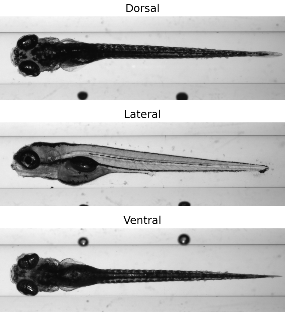
Sortivo can take 360° images of an entity with high-resolution optics. This makes it easy to image larvae from any angle without mounting them in agarose and orienting them by hand. Even with a dedicated mold, the manual mounting and orienting takes about one minute per larva (22). Once the agarose sets, the larva is fixed in a single orientation, so getting a second view means mounting it again. In Sortivo, the larva is rotated during screening, so lateral, dorsal and ventral views come from a single pass, with no manual handling. The larva is then plated directly, ready for the next step of the experiment.
The bright-field image quality is high enough to show the larva’s physiology live: the heartbeat and blood cells flowing through the vessels are visible directly in the camera feed during screening. Heart rate is a readout researchers measure in zebrafish, for example to test how small molecules affect heart function (23). Sortivo shows it without any extra preparation.
Blood Flow in a wild-type zebrafish embryos ~2dpf | Lateral | Zebrafish embryo provided by University of Fribourg.
Fluorescence Imaging
Beyond bright-field, Sortivo images larvae in fluorescence, with up to six channels. As in bright-field, the image is live: in a larva expressing a fluorescent marker in the heart, the heartbeat can be followed directly in fluorescence during screening.
GFP expression in heart | Lateral + Ventral | 1 dpi | Zebrafish embryos were provided by Monica Goldade at UZH
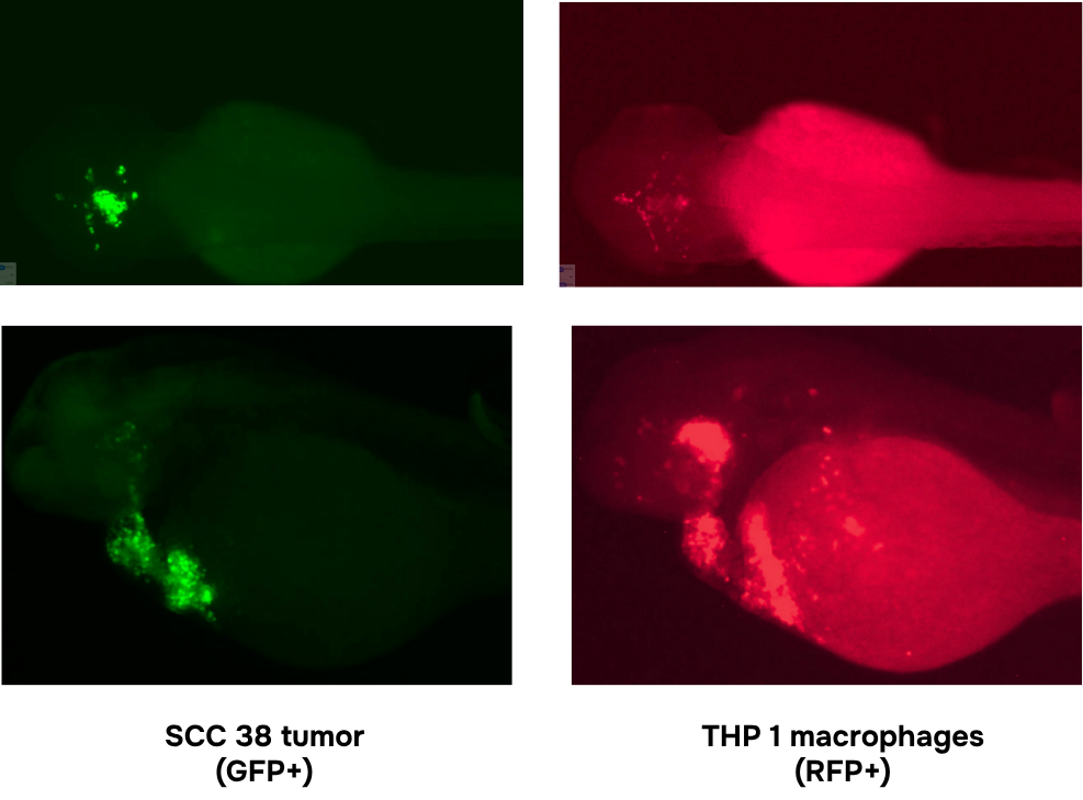
Having several channels means that several labeled cell populations can be seen in the same larva. In a xenograft, for example, human tumor cells and human macrophages carrying different fluorescent markers are imaged side by side.
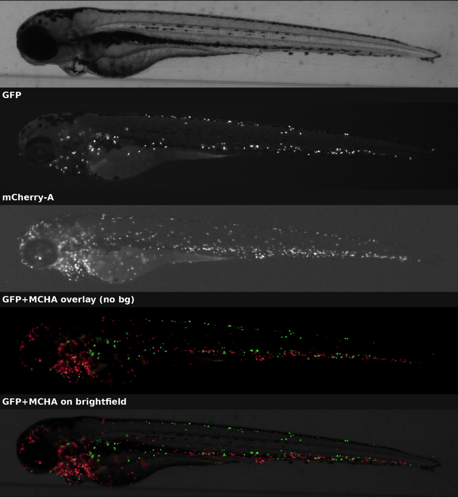
A larva is thicker than the depth of field of the camera, so a single image cannot keep every labeled cell in focus. To solve this, Sortivo’s embedded software captures z-stacks directly on the machine. It takes several images at different focus points and merges them into a single sharp image with a standard focus-stacking algorithm. No separate software or post-processing is needed, and the merged images can be combined and overlaid on the bright-field view to locate each signal in the larva.
Manual vs. Automated: How workflows change
To see what changes in practice, we compare a typical sorting and imaging routine, done by hand and with Sortivo.
(lateral, dorsal, ventral…)
By hand, the researcher does every step, one larva at a time. The larvae are first anesthetized and pre-sorted under a stereomicroscope, for example to find transgene carriers by their fluorescence (14). Each selected larva is then mounted, usually in agarose, and oriented under the microscope before it is imaged. Handling larvae for imaging is described as tedious and time-consuming (19). Even with a dedicated mold, mounting takes about one minute per larva (22).
This step becomes the bottleneck as soon as more than one view is needed. Once the agarose sets, the larva is fixed in a single orientation. A lateral view and a dorsal or ventral view therefore mean mounting, orienting and imaging the same larva again, once per view. The handling time grows with every view added. Only then is the larva recovered from the agarose and plated.
With Sortivo, this loop disappears. The researcher anesthetizes the larvae and, at the start of the run, labels a few of them as positive or negative, and Sortivo sets the sorting threshold itself. Each larva is then rotated inside the machine and imaged from all sides, in bright-field and fluorescence, in a single pass without mounting. It is sorted automatically and plated into the positive or negative output. Every larva comes out sorted, with all its views.
Conclusions and Outlooks
Sorting zebrafish larvae is an essential but time-consuming step, and imaging adds to the burden: each larva must be mounted and oriented, once for every view. Sortivo removes this loop and automates almost every other manual step. In a single run, it images each larva from all necessary angles, sorts it and plates it. The researcher only anesthetizes the larvae and labels a few examples at the start. After that, they can move on to other tasks and come back a few hours later to imaged and sorted plates.
Every larva leaves Sortivo with its images: bright-field sharp enough to follow the heartbeat and blood flow live, several fluorescence channels, and z-stacks merged on the machine. Sortivo keeps track of each larva from one plate to the next, even when the plate layout changes, so its images and data stay attached to it. The sorted plates can then become the starting point for further experiments on the same larvae.
Interested in seeing Sortivo on your own samples? Contact us to arrange a demonstration.
References
- Howe, K., Clark, M. D., Torroja, C. F., et al. (2013). The zebrafish reference genome sequence and its relationship to the human genome. Nature, 496, 498–503. https://doi.org/10.1038/nature12111
- Lieschke, G. J., & Currie, P. D. (2007). Animal models of human disease: zebrafish swim into view. Nature Reviews Genetics, 8, 353–367. https://doi.org/10.1038/nrg2091
- MacRae, C. A., & Peterson, R. T. (2015). Zebrafish as tools for drug discovery. Nature Reviews Drug Discovery, 14, 721–731. https://doi.org/10.1038/nrd4627
- Planchart, A., Mattingly, C. J., Allen, D., et al. (2016). Advancing toxicology research using in vivo high throughput toxicology with small fish models. ALTEX, 33(4), 435–452. https://doi.org/10.14573/altex.1601281
- Rennekamp, A. J., & Peterson, R. T. (2015). 15 years of zebrafish chemical screening. Current Opinion in Chemical Biology, 24, 58–70. https://doi.org/10.1016/j.cbpa.2014.10.025
- Patton, E. E., Zon, L. I., & Langenau, D. M. (2021). Zebrafish disease models in drug discovery: from preclinical modelling to clinical trials. Nature Reviews Drug Discovery, 20, 611–628. https://doi.org/10.1038/s41573-021-00210-8
- Adatto, I., Lawrence, C., Thompson, M., & Zon, L. I. (2011). A new system for the rapid collection of large numbers of developmentally staged zebrafish embryos. PLoS ONE, 6(6), e21715. https://doi.org/10.1371/journal.pone.0021715
- Lawrence, C., Ebersole, J. P., & Kesseli, R. V. (2008). Rapid growth and out-crossing promote female development in zebrafish (Danio rerio). Environmental Biology of Fishes, 81, 239–246. https://doi.org/10.1007/s10641-007-9195-8
- Nasiadka, A., & Clark, M. D. (2012). Zebrafish breeding in the laboratory environment. ILAR Journal, 53(2), 161–168. https://doi.org/10.1093/ilar.53.2.161
- Mandrell, D., Truong, L., Jephson, C., et al. (2012). Automated zebrafish chorion removal and single embryo placement: optimizing throughput of zebrafish developmental toxicity screens. Journal of Laboratory Automation, 17(1), 66–74. https://doi.org/10.1177/2211068211432197
- Pardo-Martin, C., Chang, T. Y., Koo, B. K., et al. (2010). High-throughput in vivo vertebrate screening. Nature Methods, 7, 634–636. https://doi.org/10.1038/nmeth.1481
- Parichy, D. M., Elizondo, M. R., Mills, M. G., Gordon, T. N., & Engeszer, R. E. (2009). Normal table of postembryonic zebrafish development: staging by externally visible anatomy of the living fish. Developmental Dynamics, 238, 2975–3015. https://doi.org/10.1002/dvdy.22113
- Truong, L., Reif, D. M., St Mary, L., et al. (2014). Multidimensional in vivo hazard assessment using zebrafish. Toxicological Sciences, 137(1), 212–233. https://doi.org/10.1093/toxsci/kft235
- Kwan, K. M., Fujimoto, E., Grabher, C., et al. (2007). The Tol2kit: a multisite gateway-based construction kit for Tol2 transposon transgenesis constructs. Developmental Dynamics, 236, 3088–3099. https://doi.org/10.1002/dvdy.21343
- Diouf, A., Sadak, F., Bereziat, L., et al. (2025). Combining deep learning and microfluidics for fast and noninvasive sorting of zebrafish embryo. Scientific Reports, 15, 37778. https://doi.org/10.1038/s41598-025-17946-7
- Saint-Amant, L., & Drapeau, P. (1998). Time course of the development of motor behaviors in the zebrafish embryo. Journal of Neurobiology, 37(4), 622–632. https://doi.org/10.1002/(SICI)1097-4695(199812)37:4<622::AID-NEU10>3.0.CO;2-S
- Teixidó, E., Kießling, T. R., Krupp, E., et al. (2019). Automated morphological feature assessment for zebrafish embryo developmental toxicity screens. Toxicological Sciences, 167(2), 438–449. https://doi.org/10.1093/toxsci/kfy250
- Jain, G., & Shetty, P. (2014). Occupational concerns associated with regular use of microscope. International Journal of Occupational Medicine and Environmental Health, 27(4), 591–598. https://doi.org/10.2478/s13382-014-0288-2
- Pulak, R. (2016). Tools for automating the imaging of zebrafish larvae. Methods, 96, 118–126. https://doi.org/10.1016/j.ymeth.2015.11.021
- Cani, O., Zannotti, L., De Vita, A., et al. (2026). Zebrafish tumor xenograft models for drug-screening: a systematic review of methods for treatment assessment. Journal of Translational Medicine, 24, 417. https://doi.org/10.1186/s12967-026-07787-x
- Ding, Y., van der Kolk, K. J., van der Ent, W., et al. (2025). Automated microinjection for zebrafish xenograft models. npj Biomedical Innovations, 2, 13. https://doi.org/10.1038/s44385-025-00016-y
- Geng, Y., & Peterson, R. T. (2021). Rapid mounting of zebrafish larvae for brain imaging. Zebrafish, 18(6), 376–379. https://doi.org/10.1089/zeb.2021.0062
- Burns, C. G., Milan, D. J., Grande, E. J., Rottbauer, W., MacRae, C. A., & Fishman, M. C. (2005). High-throughput assay for small molecules that modulate zebrafish embryonic heart rate. Nature Chemical Biology, 1(5), 263–264. https://doi.org/10.1038/nchembio732